 Extracts of the MLs have been utilized for traditional medicines to cure diabetes, bronchitis, diarrhea, asthma, kidney, scabies, respiratory problems, syphilis, and urinary disorders [2,3]. The most active biological constituent of MLs is mangiferin, followed by phenolic acids, benzophenones, and other antioxidants such as flavonoids, carotenoids, quercetin, isoquercetin, ascorbic acid, and tocopherols. Mangiferin is the main contributor of most of the biological activities of MLs extract. MLs have a great scope of valorization as they are recognized to possess varied phytochemical, biological, and pharmacological properties, viz. anti-microbial, antioxidant, anti-diabetic, anti-tumour, and immunomodulatory effects. ML oil (MLO) contains monoterpenes, sesquiterpenes, minor quantities of other analogues, and trace amounts of non-terpenoid hydrocarbons and oxygenated hydrocarbons. The essential oil from MLs also possesses bacteriostatic properties and contains several antimicrobial constituents such as α-gurjunene, trans-caryophyllene, α-humulene, α-selinene, and camphor. The benzophenone derivatives in MLs possess significant α-glucosidase inhibitory and immunosuppressive activities. There are several reviews that have been developed to discuss the bioactive compounds and health promoting effect of mango fruit/pulp [4,5,6,7], whereas others contain a scattered compilation of literature on mango seeds, MLs, and mango bark [8,9].
Anti-Diabetic Activity
Diabetes is a chronic metabolic disorder that badly disturbs the health and quality of human life and is established as the foremost threat to society irrespective of geographical locations. Diabetes is characterized by elevated glucose or above-normal glucose level (70–110 mg/dL), which are partially due to oxidative damage to pancreatic β-cells, leading to a decline in insulin secretion [44]. Insulin regulates the blood glucose level (BGL); low secretion of insulin causes hyperglycemia, which enhances oxidative stresses and eventually causes several health problems like frequent urination, thirst, and hunger [45]. In 2016, the International Diabetes Federation (IDF) reported that around 415 million people are diabetic, with a population of 642 million predicted to suffer from type-2 diabetes (diabetes mellitus or DM) by 2040 [46]. Several medicines such as acarbose are currently used in diabetes, but such types of diabetic medicines lack DM restraint and revealed undesirable side effects over time [45]. All over the world, researchers are exploring medicinal plants as an effective way to cure this debilitating disorder, because medicinal plants are a rich source of bioactive constituents, and most of them are known to be potent against DM.
MLs have been widely claimed as effective ethnomedicine against DM due to their anti-diabetic bioactive constituents like benzophenones (mangiferin) and flavonoids (quercetin and its glucoside forms). One of the best effective approaches in the cure of DM is the inhibition of α-amylase and α-glucosidase enzymes, which regulate postprandial glucose absorption [47]. A comparative analysis of mangiferin and MLs extract was done to check the efficiency of each extract to inhibit α-glucosidase enzymes. MLs extracts at a concentration of 100, 250, and 500 mg/mL caused up to 77.8%, 83.4%, and 95.7% inhibition of α-glucosidase, respectively. At the same time, mangiferin at a concentration of 10, 25, and 50 resulted in 86.85%, 92.35%, and 99.11% inhibition of α-glucosidase, respectively. It can be inferred that mangiferin is an active ingredient in the inhibition of α-glucosidase enzyme activity and in managing the diabetic condition [48]. Ganogpichayagrai et al. [42] evaluated the anti-diabetic activity of mangiferin and MLs extract through the inhibition of α-glucosidase and α-amylase in vitro. Authors reported that mangiferin showed strong inhibition of rat α-glucosidase with a median inhibitory concentration IC50 of 433.3 µg/mL and MLs extract showed potent inhibition of yeast α-glucosidase with the IC50 of 50.3 µg/mL. Saleem et al. [49] evaluated the anti-diabetic potential of MLs extract (550, 750, 950 mg/kg) cv. Ratol in alloxan monohydrate (150 mg/kg) induced diabetes in albino mice. Authors found that the administration of MLs extract in chemically induced diabetic mice reduced the postprandial glucose level, prevented the surge of glucose in the blood, and improved the lipid profile along with body weight. Different bioactive compounds have been isolated from the MLs extract and demonstrated their anti-diabetic potential. Gu et al. [31] isolated and characterized four bioactive compounds as manindicins A and B, mangiferin, and norathyriol (deglycosylated mangiferin) from MLs extract. Authors revealed that norathyriol exhibited strong α-glucosidase inhibition with IC50 of 4.22 ± 0.19 μg/mL, which was four-fold effective with respect to commercial inhibitor acarbose (IC50: 16.28 ± 1.22 μg/mL), while mangiferin (IC50: 32.11 ± 2.01 μg/mL) and manindicin A (IC50: >300 μg/mL) and B (>300 μg/mL) displayed weaker α-glucosidase inhibition. The less inhibitory potential of Mangiferin may be due to its molecular size and polarity. The replacement of glucose moiety with hydrogen may weaken the steric hindrance during mangiferin–enzyme interaction and enhance the inhibitory potential of mangiferin towards α-glucosidase [31]. The anti-diabetic potential of the mangiferin is schematically shown in Figure 3. The anti-diabetic potential of mangiferin was also demonstrated, as it increases insulin sensitivity and inhibits α-glucosidase [50]. Similarly, for quercetin, α-glucosidase inhibition was substantially higher than that of its 3-O-glucoside hyperoside [51]. Bhuvaneshwari et al. [52] investigated the anti-diabetic activity of tender and mature leaves of totapuri cultivar of mango, and authors found that tender leaves extract (500 mg/kg) efficiently inhibited the α-amylase with IC50 22.01 µg/mL, while mature leaf extract (500 mg/kg) exhibited the α-glucosidase inhibition with IC50 21.03 µg/mL. Findings suggest that bioactive compounds from the ML can be effective in reducing the risk of diabetes.

Schematic showing anti-diabetic activity of the mangiferin from mango leaves.
Antioxidant Activities
Many recent studies have shown that free radicals generated during the metabolic process contribute to various degenerative diseases such as acquired immunodeficiency syndrome, ischaemic diseases, neurological disorders, and many more [53]. Antioxidant substances, on the other side, provide a high level of antioxidant activity to lessen the adverse effects of free radicals. MLs were reported to have antioxidant capacity due to the presence of phenolics and flavonoids in different studies [54]. High-performance liquid chromatography coupled to electrospray ionization and quadrupole time-of-flight mass spectrometry (HPLC-ESI-qTOF-MS/MS) analysis of MLs extract had identified neomangiferin, mangiferin, kaempferol-3-O-rutinoside, isoquercitrin, and quercetin as the main compounds and also reported that these compounds contributed directly to the antioxidant activity of MLs [55]. The 2,2-diphenyl-1-picrylhydrazyl (DPPH) assay and superoxide dismutase (SOD)-like activity had shown that MLs serve as a moderate antioxidant with an IC50 value of ~9 and 117 μg/mL [56]. In another analysis, MLs methanol extract provided radical scavenging activity with an IC50 value of 13.37 μg/mL [57]. Fraction analysis of MLs extract with n-butanol, hexane, and ethylacetate demonstrated that ethylacetate fraction had the highest antioxidant capacity of 1226 and 2817.99 mg trilox/g estimated using DPPH and 2,2′-azino-bis-3-ethylbenzthiazoline-6-sulphonic acid (ABTS) assay, respectively, and reducing power of 10172.59 µmol FeSO4.7H2O/g extracts analyzed through ferric reducing antioxidant power (FRAP) assay [58]. Similarly, subcritical water extracts of MLs had antioxidant activity index (AAI) values of 7.92 ± 0.16 and demonstrated superior activity to (+)-α-tocopherol (AAI = 3.65 ± 0.07) [41]. Trolox equivalent antioxidant ability (TEAC) study of MLs extracts had recorded 2.13 and 2.59 mmol TE/g DW TEAC values, respectively, for mangiferin pentoside and benzophenones [59]. The efficacy of the MLs was also studied in the chitosan-based films, and it was found that antioxidant capacity of the MLs supplemented chitosan films improved in a dose-dependent manner [60]. The antioxidant activity of hydroalcoholic MLs extract fermented with either Lactobacillus casei or effective microorganisms had higher antioxidant activity. The study also showed that fermented extracts decreased lipopolysaccharide-generated reactive oxygen species [61]. In an advanced study, MLs extract was found suitable as a green antioxidant for increasing the storage life of biodiesel [62]. In summary, many interesting results indicated the potential of MLs extract as an antioxidant with wider applicability in food, food packaging, and many more industries.
Antimicrobial Activities
There is immense interest in unravelling the role of bioactive compounds present in nature. Some medicinal plants with antimicrobial attributes are capable of evading the activity of multi-drug resistant (MDR) microbes, which helps in withstanding antimicrobial resistance [63]. Distinct morphological parts of the mango plant like leaves, stem, kernel, seeds, and bark have been manifested to show antimicrobial activities against microbes like Staphylococcus sp., Bacillus subtilis, Escherichia coli, Candida albicans, Proteus vulgaris, Pseudomonas fluorescens, Shigella flexneri, Klebsiella pneumoniae, and Salmonella typhi. MLs extract is the most studied part for antibacterial effects. Bharti [64] observed that hexane and hexane/ethyl acetate extracts of MLs exhibit favorable antibacterial effects against Mycobacterium tuberculosis and Enterobacter aerogenes. Antimicrobial investigation of the essential oils extracted from leaves of five Egyptian mango cultivars to be used as preservatives materials has been demonstrated by Ouf et al. [18] against Staphylococcus sp. (Minimum Inhibitory Concentration (MIC): 62.5 μg/mL), Bacillus subtilis (MIC: 125 μg/mL), Escherichia coli (MIC: 125 μg/mL), Pseudomonas aeruginosa (MIC: 500 μg/mL), Aspergillus flavus (MIC: 1000 μg/mL), and Salmonella typhi (MIC: 1000 μg/mL). The major phytochemicals responsible for the antimicrobial activity in mango leaves include phenolics, alkaloids, saponins, glycosides, terpenes, and tannins. The concentration of the aforementioned compounds were measured as follows: flavonoid content was the highest at 11.25 mg/100 g; there was 3.23 mg/100 g of saponins; phenolic content was 0.08 mg/100 g; and tannins in leaves was at 0.46 mg/100 g [24]. Polyphenols and phenolic acids present in ML extract include protocatechuic acid, gallic acid, hyperin, catechin, quercetin, kainic acid, ethyl digallate, ellagic acid, and shikimic acid, which can inhibit the growth of pathogens [8]. The mechanism of exertion of antimicrobial activity by these compounds involves depleting intracellular ATP levels, depolarization of plasma membrane, cytoplasm leakage, damaging genetic material, and declining the concentration of microbial protein [65]. Anti-microbial activity of mangiferin is schematized in Figure 4. Additionally, an adequate level of antibacterial activity of leaf extract was found against Gram positive bacteria, but no or less activity against Gram-negative bacteria was observed [66]. The study indicated that MLs extract exhibited diameter of zone of inhibition in the range of 7.0‒11.5 mm against Gram-positive bacteria like Staphylococcus and Bacillus sp., but no activity was seen against gram-negative Salmonella spp. Additionally, Mangiferin, a xanthone C-glycosyl compound extracted from MLs extract, has also shown to possess strong iron chelating activity, which favors antimicrobial activity. Furthermore, chemical analysis of MLs extract for antimicrobial activity indicated the presence of five major flavonoid compounds including epicatechin-3-O-β-glucopyranoside, 5-hydroxy-3-(4-hydroxyl phenyl) pyrano chromene-4 (8H)-one, 6-(phydroxybenzyl) taxifolin-7-O-β-D-glucoside, quercetin-3-O-α-glucopyranosyl-(1-2)-β-D-glucopyranoside, and epicatechin(2-(3,4dihydroxyphenyl)-3,4-dihydro-2H-chromene-3,5,7-triol [67]. These compounds are identified to be synthesized immediately after a fungal attack, and a concentration of 1000 ppm has curtailed the growth of target fungal species like Aspergillus and Alternaria from 56‒97% [67]. A myriad of terpenes identified from MLs extract including α-pinene, β-pinene, δ-elemene, taraxerol, β-elemene, camphene, γ–cadinene, lupeol, friedelin, linalool, α-guaiene, humulene, α-farnesene, myrcene, limonene, β-ocimene, γ–terpinene, and α-terpinolene exhibit bacteriostatic and bactericidal effects against different pathogens [8]. HPLC-TOF-ESI/MS analysis of leaf extract to identify hydrolyzable tannins revealed the presence of gallatotannins. Antimicrobial properties of gallatotannins have been associated with their ability to hinder the enzymes of the pathogen, disintegrate lipid bilayer membranes, and promote chelation of metal ions [68]. The MIC of MLs extract is reported to be in the range 11–52 mg/mL [69] and the MIC values were greatest against E. coli with 36.3 mg/mL. The aforementioned phytocompounds purified from ML extract can be directly used as food additives to enhance the shelf-life of foods, and as an alternative for the synthetic antimicrobial agents, owing to their broad biological and pharmacological activities [70].
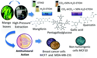
Mango leaves and cancer
Anticancer Activities
Cancer is one of the most prevalent global threats after cardiovascular disease. Thus, there is an imperative need to undertake novel treatment strategies to counter this global issue. Polyphenols present in MLs like gallotannins, phenolic acids, quercetin, and mangiferin exhibit chemo-preventive effects against various cancer types due to their anti-inflammatory and antioxidant effects [35]. Antitumoral activities of MLs extract are mainly attributed to the primary bioactive xanthone glucoside, mangiferin. These compounds are demonstrated to suppress several cancers by impeding their invasion, migration, and proliferation [36]. Mangiferin is found to overturn the transition from epithelial-to-mesenchymal in MCF7 breast cancer cell lines by inhibiting Wnt/β-catenin pathway and by downregulating the expression of specific enzymes (5′Nucleotidase, γ-GT, and aryl hydrocarbon hydroxylase) in lung cancer bearing albino mice [37,38]. It also suppresses the levels of Akt phosphorylation and cyclin B1, causing spontaneous cell cycle arrest in G2/M phase. It was also found to instigate Nrf2-mediated antioxidant activities at a concentration of 50 μM, with no influence of myeloid leukaemia cell sensitivity to chemotherapeutics [39]. Mangiferin was also found to mitigate the oxidative stress and inhibit methylmercury-induced DNA damage in human neuroblastoma cell line IMR-32 [40]. The anti-invasive and antimetastatic activities of mangiferin could also be attributed to its ability to regulate the expression of metalloproteinases, which determines the cell proliferation and inhibits epithelial–mesenchymal transition, eventually causing a loss in cell adhesion.
A study was conducted to investigate the antitumoral effects of ML extracts on (MDA-MB-231) highly and (MCF7) minimally invasive breast cancer cells and (MCF10) non-tumorigenic cells at IC50 >200 µg/mL [41]. The leaf extracts displayed protective properties against cytotoxic and oxidation effects on breast cancer cell lines and minimal damage to non-carcinogenic cells. MLs extracts with a high concentration of homo-mangiferin and methyl gallate were found more effective against MDA-MB-231 cells, while gallotannins showed cytotoxicity against MCF7 cells. In another study, ethanolic extract of mango leaves at a concentration of IC50 >200 µg/mL exhibited cytotoxic activities against lung fibroblast (ATCC CLS 300421,WI-38 VA-13 subline 2RA), skin fibroblast (ATCC CRL1947, CCD-986SK), colon adenocarcinoma (ATCC CCL227, SW 620), gastric carcinoma (ATCC HTB103, Kato-III), liver hepatoblastoma (ATCC HB8065, Hep-G2), bronchogenic carcinoma (ATCC HTB-168TB, Chago K-1), and ductal carcinoma (ATCC HTB20, BT474) [42]. Similarly, MLs extract was used to synthesize silver nanorods. These nanorods exhibited strong in vitro cytotoxicity, antioxidant, and anticancer activities at 10% w/v against colorectal carcinoma and breast cancer cell lines (HCT-116, MCF-7) [43]. Anti-cancer potential of the mangiferin is schematically depicted in Figure 2.

Schematic showing anti-cancer activity of the mangiferin from mango leaves.